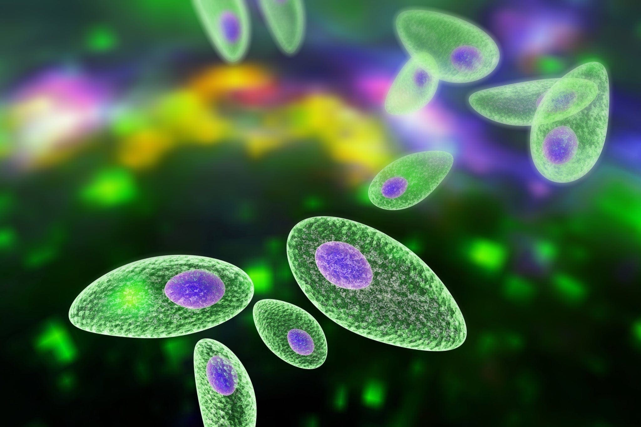
USMLE Toxoplasmosis (TORCH infection)
 [1]
[1]Risk Category: Pregnant women; debilitated patients. If HIV positive CD4 count less than 100
Mode of Transmission:
1) Oocysts in cat feces
2) Contaminated meat having oocysts
3) Through placenta (if mother acquires Toxoplasmosis during pregnancy)
Imaging: (CT/MRI)
Ring enhancing lesion
Triad:
1) Intracranial calcification (Dystrophic calcification)
2) Hydrocephalous
3) Chorioretinitis
Treatment:
Sulfadiazine plus Pyrimethamine
Prophylaxis in AIDS patient:
Trimethoprim and Sulfamethoxazole
References:
[1] https://familydoctor.org/condition/toxoplasmosis/ [Online] / auth. familydoctor.org // https://familydoctor.org.
Internal Iliac Artery
Common Iliac Artery bifurcates at the level of L4; into External and Internal Iliac artery. External Iliac artery provides blood supply into peripheral areas via several other small branches

Internal Iliac artery plays a vital role in supplying pelvis and pelvic viscera.
Internal Iliac Artery further branches into Anterior and Posterior Branches at the upper margin of greater sciatic foramen; smaller than external iliac artery.
Relation of Internal Iliac artery: [2]
Posterior to the ureter
Anterior to the internal iliac vein, lumbosacral vein and piriformis muscle.
Branches of Internal Iliac artery:
A) Anterior Branches: can be memorized as 3 Urinary Branches. 3Visceral branches & 3superficial branches. [3]
a) 3 Urinary Branches:
i) Superior vesicle artery
ii) Inferior vesicle artery
iii) Umbilical artery (patent only in fetus)
b) 3 Visceral branches:
i) Uterine artery
ii) Vaginal artery
iii) Middle Rectal artery
c) 3 superficial branches
i) Internal Pudendal artery
ii) Obturator artery
iii) Inferior gluteal artery
B) Posterior Branches: can be memorized as IL-LS-SG
a) IL: Ilio-lumbar artery
b) LS: Lumbo-sacral artery
c) SG : Superior gluteal artery (contrast Inferior Gluteal artery in Anterior branch)
Diagram: [1]

References:
[1] Drake, R. L., Vogl, W., & Mitchell, A. W. (2014). Gray’s Atlas of Anatomy. Elsevier.
[2] Wikipedia.org . (n.d.). https://en.wikipedia.org/wiki/Internal_iliac_artery. Retrieved from https://en.wikipedia.org.
[3] Yang , R., & Bashir , O. (n.d.). https://radiopaedia.org/articles/internal-iliac-artery. Retrieved from https://radiopaedia.org.
Medicine would Never have been the same without them!
-Karun Bhattarai
MBBS BPKIHS (Batch 2018)

The Doctor Who Drank Infectious Broth, Gave Himself an Ulcer, and Solved a Medical Mystery!
In 1910 an article was published in the
Lancet stating "acid gastritis" or “hyperchlorhydria” as the sole
cause for Duodenal Ulcer (1) . Therefore, in order
to treat duodenal ulcer, truncal vagotomy or antrectomy (cut off the bottom of
the stomach and reconnect to the intestine) was considered as the only
treatment available for about seven decades. Marshall while he was in the third
year of his internal medicine training, in 1981; worked on a project with Dr.
Robin Warren, the hospital pathologist, who was seeing some bacteria on
biopsies of all ulcer and stomach cancer patients for two years, which were all
identical. Microbiologists had no dogma to overcome about the causes of
gastritis and peptic ulcers but the wider medical community remained hard to
convince. Dr Marshall had a patient with gastritis. He got the bacteria and
cultured them, then worked out which antibiotics could kill patient’s infection
in the lab (in that case, bismuth plus metronidazole). He treated the patient
and did an endoscopy to make sure his infection was gone. The same year, in an
act born to some extent of frustration, Marshall deliberately infected himself
by drinking a solution swimming with the bacterium, as part of a successful and
widely reported experiment to prove Koch's postulates. But many clinicians
still remained unmoved. It wasn't until the early 1990s that the evidence of
Marshall and Warren became impossible to ignore, at which point pharmaceutical
development and clinical practice underwent a shift towards eradication of H.
pylori to treat ulcers. For their discovery of the bacterium Helicobacter
pylori and its role in gastritis and peptic ulcer disease, the Nobel Prize in
Physiology or Medicine 2005 was awarded jointly to Barry J. Marshall and J.
Robin Warren (2) .
Medical Imaging Technology (CT Scan,MRI) and Non-Interventional Visualization of Body!
One of the England engineer Godfrey
Hounsfield came up with an idea that instead of taking x-ray from just one
angle, if taken from all angles around one could determine something covered
inside the box. He then set to work constructing a computer that could take
input from X-rays at various angles to create an image of the object in
"slices". In 1979 Cormack A.M
and Hounsfield G.N were jointly awarded the Nobel Prize in Physiology or
Medicine for the development of computer assisted tomography (CAT), popularly
known as CT Scan these days. But it came with great bane of radiation
hazard!
Continuous efforts were being done in
advancement of imaging technology where the principle of Nuclear Magnetic
Resonance (Felix Bloch and Edward Purcell, 1946 NP1952) originated but was not
kept in clinical practice yet. Up until 1970s MRI was being used just for
chemical and physical analysis of molecules. Early detection of internal
neoplasm was greatly hampered during those days because of increased
permeability of many tumors to x-rays. A study found different biological
responses among Tumor cells and Normal cells when resonated magnetically within
magnetic field (3) . Consequently, the mankind was able to scan
human anatomy without a drop of radiation using a different principle than that
of CT scan. Instead of x-rays field used in CT Scan, MRI used Magnetic field to
resonate the protons present in fats and water in human body and using complex
algorithms transcribed into the series of images of scanned section. In
recognition of this contribution to mankind, the Nobel Prize in Physiology or
Medicine of 2003 was awarded jointly to Paul C. Lauterbur and Sir Peter
Mansfield "for their discoveries concerning magnetic resonance imaging (MRI)."
Project 523; Artemisinin yielding ancient Herbs and Nobel Prize
According to a recent WHO report, 97
countries have ongoing malaria transmission, and an estimated 3.4 billion
people are at risk of malaria, of whom ~1.2 billion are at high risk (4) .
What if the secret of modern medicine is encrypted
within the ancient herbs? It was early 1970s in China (period when scientific
research was strictly prohibited). In response to a request from the Vietnam
government for help on malaria treatment, the Chinese government launched a
secret operation called 523 Project during their Cultural Revolution. Professor
Youyou Tu (an 84-year-old, female scientist) joined the project; she searched
more than 2,000 recipes and compiled 640 recipes used for the treatment of
fever written ~1700 years ago. Professor Tu suddenly realized that high
temperature could be the cause of instability in antimalarial activity they
experienced. She decided to use ether, replacing ethanol, to extract the active
ingredients from the plant leaves. Professor Tu was the person who discovered
an efficient method for extracting the active ingredient from the A. annua plant (5) (the plant which the
world was witnessing for years and yet was left unnoticed!)
For her discoveries concerning a novel
therapy against Malaria, Prof Youyou Tu shared Nobel Prize for Medicine and
Physiology in 2015.
References
1. Some points in the diagnosis and treatment of
chronic duodenal ulcer. Moynihan, B.G.A. 4610, 1912, The Lancet,
Vol. 179, pp. 9-12.
2. Nobel Prize
winners Robin Warren and Barry Marshall. The Lancet . 9495,
s.l. : Elsevier Ltd., 10 22, 2005, The Lancet , Vol. 366, p. 1429.
3. Tumor Detection
by Nuclear Magnetic Resonance. Damadian, Raymond. Mar 19, 1971,
Science, pp. 1151-1153.
4. World Health
Organization. World Malaria Report 2014. s.l. : WHO , 2014.
p. 3. 978 92 4 156483 0 .
5. The discovery
of artemisinin and the Nobel Prize in Physiology or Medicine. SU
Xin-Zhuan, MILLER Louis H. 11, s.l. : Springer , 10 16, 2015,
SCIENCE CHINA Life Sciences, Vol. 58, pp. 1175–1179.
Kerley A,B and C Signs Chest X-ray
In patients with INTERSTITIAL LUNGS Disease, fluid or cellular infiltration of inflammatory (edematous) fluids into the interstitium of lungs causes thin pulmonary opacity; this presents a radiographic opacity along the alveoli, known as Kerley Sign.
Behind the Scene:
[A] Cardiac
a. Congestive Heart Failure( Left Sided Heart Failure)
[B] Extracardiac
a. pulmonary fibrosis
b. interstitial deposition of heavy metal particles (Pneumoconiosis)
c. carcinomatosis of the lung
TYPES OF KERLEY SIGNS
Kerley A lines
These signs present as a diagonal lines with its course from the hilum of the lungs and extends out to the periphery of the lungs which is caused because of the anastomotic channel distention between the central lymphatics of the lungs and the peripheral draining lymph vessels. Kerley A sign is a must when Kerley B and/or C lines are seen. However, this sign itself is quite rare.
Kerley B lines
Kerley B sign present as a short parallel lines across the outward pheripheral zone of the lungs along the pleura. Those interlobar septated area are right angle to the pleura but are absent along the fissure and are most commonly reported from the zone III of lungs along Costophrenic angle(PA View) and in substernal region on lateral view.
Causes:
i. pulmonary edema
ii. lymphangitis carcinomatosa
iii. malignant lymphoma
iv. viral and mycoplasmal pneumonia
v. interstitial pulmonary fibrosis
vi. pneumoconiosis
vii. sarcoidosis
They can be an evanescent sign on the chest x-ray of a patient in and out of heart failure.
Kerley C lines
Kerley C lines are most rare. These lines present as short and fine lines with reticular appearance along the lungs parenchyma.
Question Bank for Acadeimc Year 2019

Question Bank First Year =====> Question Bank First Year 2019
Question Bank Second Year ===> Question Bank Second Year 2019

Radiographic Equipment B.Sc. MIT Year-1 IOM (MMC) Internal Assessment 2075
Bachelor Level – B.Sc. MIT / First Year/ IOM Time:- 45 Min.
Radiographic Equipment IV FM :
Q.1 Explain the features of cathode assembly of a rotating anode x-ray tube.
Q.2 What is line focus principle and anode heel effect?
Q.3 What is photoelectric effect and what are its significance in diagnostic radiology?
Q.4 Explain the construction and function of a linear x-ray grid?
Q.5 What is an automatic exposure timer? Explain its working principle.
Q.6 How is kilovoltage (kV) controlled in an x-ray circuit?
Human Physiology B.Sc. MIT Year-1 IOM (MMC) Internal Assessment 2075
Bachelor Level – B.Sc. MIT / First Year/ IOM Time:- 60 Min.
Human Physiology II FM : 35
Q.1 Describe the actions of insulin. Explain the features of hypoglycemia and diabetes mellitus. (6+2+2)
Q.2 Define synapse and briefly describe the events involved in the synaptic transmission. (1+4)
Q.3 Define glomerular filtration rate. Explain any TWO factors affection GFR. (1+2+2)
Q.4 Give the composition and functions of gastric juice. (2+3)
Q.5 Explain the functions of inner ear. Add a note on deafness. (3+2)
Q.6 List any four gastro-intestinal hormones. Give the actions of each. (1+4)
Human Physiology Pre-Test B.Sc. MIT Year-1 IOM (MMC) Internal Assessment 2075
Bachelor Level – B.Sc. MIT / First Year/ IOM Time:- 60 Min.
Human Physiology II FM : 35
Q.1 Name the different types of transport mechanisms across the cell membrane. Explain any TWO of them giving appropriate examples. (2+3)
Q.2 Name the blood group systems. Describe the indications and hazards of blood transfusion. (2+4+4)
Q.3 Define cardiac output. Explain the factors regulating cardiac output. (1+4)
Q.4 Define vital capacity. Give the normal values in adult males and adult females. Mention any ONE condition each that increase and decrease the vital capacity. (1+1+1+1+1)
Q.5 Classify muscles. Give any four functional difference between skeletal muscle and smooth muscle. (1+4)
Q.6 Name the in vivo and in vitro anticoagulants. Explain the mechanism of action of any two anticoagulants. (2+3)
Radiographic Technique B.Sc. MIT Year-1 IOM (MMC) Internal Assessment 2075
Bachelor Level – B.Sc. MIT / First Year/ IOM Time:- 90 Min.
Radiographic Technique VI FM : 40
Q.1 Write in detail about the indications, patient preparation and centering ray for chest radiography. (10)
Q.2 Write the technique of lumber x-ray AP and Lateral views. (10)
Q.3 Describe the technique for cervical spine oblique views. (10)
Q.4 Describe the technique for skull x-ray PA and Towne's view. (10)
Q.5 Write the center point of the following x-ray views: (2*5)
a. Skull Lateral view
b. Single Hip joint AP view
c. Lumber spine lateral view
d. calcaneum axial view
Human Physiology BPH Year-1 IOM (MMC) Internal Assessment 2075
Bachelor Level – BPH / First Year/ IOM Time:- 60 Min.
Human Physiology II FM : 25
Attempt any five questions.
Q.1 What is total body water? Explain how total body water is classifed into various fluid compartments. (1+4)
Q.2 Define blood pressure. Mention normal range of systolic and diastolic blood pressure in adult male. Explain the factors affecting blood pressure. (1+1+3)
Q.3 Name the different plasma proteins in blood and describe their functions. (1+4)
Q.4 What are the different types of emphysema? Write different between them. (2+3)
Q.5 Define tidal volume, vital capacity and timed vital capacity. Mention their normal values. (1.5+1.5+1.5+0.5)
Q.6 Define jaundice. Name different types of jaundice based on aetiology. (2+1+1+1)
Q.7 List the functions of secretions of oral cavity, stomach and pancreas. (5)
Q.8 Explain the physiological basis of blood groups. Mention the dangers of mismatched blood transfusion. (3+2)
Q.9 Define cardiac output, coronary artery disease, heart failure, Rheumatic heart disease and atherosclerosis. (5)
Radiographic Photography B.Sc. MIT Year-1 IOM (MMC) Internal Assessment 2075
Bachelor Level – B.Sc. MIT / First Year/ IOM Time:- 45 Min.
Radiographic Photography V FM : 80
Q.1 What is radiographic image? How radiographic image is formed according to Gurney Mott's theory? (20)
Q.2 What is intensifying screen? Describe the construction of intensifying screen with diagram and its uses. (20)
Q.3 What is characteristics curve? How they are formed? Describe it with diagram and its uses. (20)
Q.4 Write short notes on: (4*5)
a. Radiographic cassette
b. Steps of film processing(conventional)
c. Dry film technology(modern concept)
d. Exposure factors used in diagnostic radiography
B.Sc. MIT Year-2 IOM (MMC) Board Exam 2072 Physics of Modern Imaging Technology
Bachelor Level – B.Sc. MIT / Second Year/ IOM Time:- 3hrs.
Physics of Modern Imaging Technology XII FM :80
1. Discuss the importance of image matrix and pixel bit depth in digital imaging. List the advantages of digital imaging. (10) 2. Explain the working principle of helical CT and discuss the detector configuration and their construction in MDCT. (10) 3. What are slip rings in CT? Discuss types and functions of slip rings used in CT. (10) 4. Discuss how T1 and T2 weighted images are created in spin echo method of MR Imaging. (10) 5. Explain how gradient echo method of MRI is faster than spin echo MRI. (10) 6. Explain 5 common artifacts in CT images. (10) 7. Explain briefly:(5+5) a. Direct digital radiography b. Gradient coils in MRI 8. Define the following terminologies. (2*5) a. CT number b. Repetition time (TR) c. Slew rate d. Numerical size in digital image e. CTDI |
B.Sc. MIT Year-2 IOM (MMC) Board Exam 2073 Physics of Modern Imaging Technology
Bachelor Level – B.Sc. MIT / Second Year/ IOM Time:- 3hrs.
Physics of Modern Imaging Technology XII FM :80
Q.1. What is digital imaging? Discuss the important features and advantages of digital imaging. (10) Q.2. Explain the working principle of computed radiography (CR), direct radiography (DR), and digital fluoroscopy(DF). (10) Q.3. Explain the working principle of MDCT and its advantages & challenges. Discuss the significance of pitch in helical CT. (5+5) Q.4. Explain spin echo method of MR Imaging. Discuss the basic spin echo-based signal suppression technique. (10) Q.5. Explain five types of common artifacts generated in CT and in MRI. (10) Q.6. Write short notes on: (5+5) a. Slip ring technology b. X-ray tubes used in CT Q.7. Explain briefly:(5+5) a. PACs b. MRI magnets Q.8. Define the following terminologies. (2*5) a. PET-CT b. Longitudinal relaxation and T1 time c. Transverse relaxation and T2 time d. Acoustic impedance e. Doppler shift |


B.Sc. MIT Year-2 IOM (MMC) Board Exam 2074 Physics of Modern Imaging Technology
Bachelor Level – B.Sc. MIT / Second Year/ IOM Time:- 3hrs.
Physics of Modern Imaging Technology XII FM :80
| Q.1 Explain the important features of a digital image. Discuss direct and indirect conversion digital radiography in brief. (5+5) Q.2. Explain the working principle of helical CT. Discuss the features of modern MDCT. (5+5) Q. 3. Explain 3 types of artifacts in CT and MR imaging each. (10) Q. 4. What are different types of ultrasound transducers? Explain electronic transducers. (4+6) Q. 5. What is T1 and T2 relaxation in MRI? Explain T1 and T2 weighted contrast in MRI? (4+6) Q. 6. What are different methods of MR imaging? Explain basic gradient echo pulse sequence. (2+8) Q. 7. Explain briefly: (5+5) a. Pitch in helical CT b. Gamma camera Q.8. Write short notes on: (5+5) a. Gradient coils in MRI b. PACs |
Subscribe to:
Posts
(
Atom
)